Aorta Anatomy Isthmus
247 040 cm.

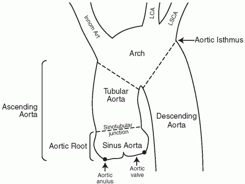
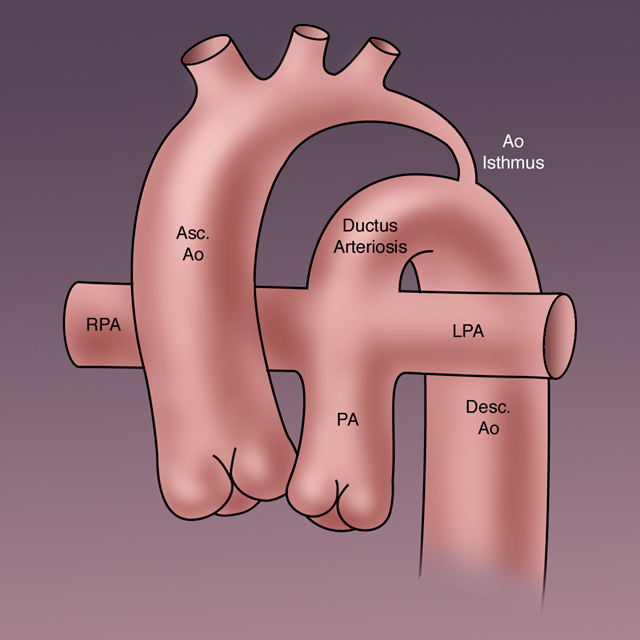
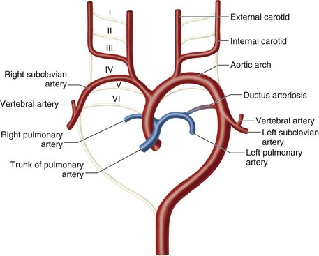
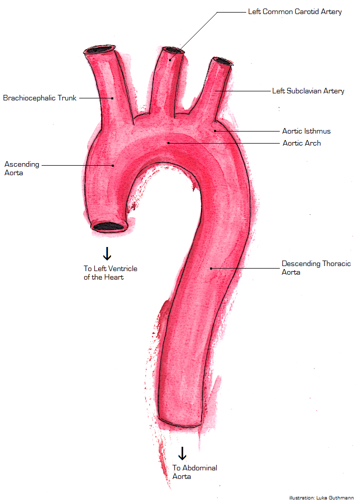
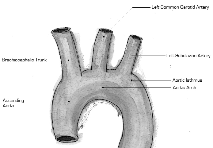
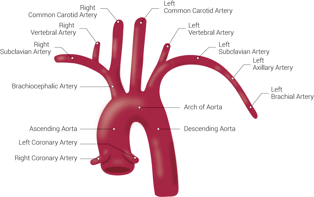
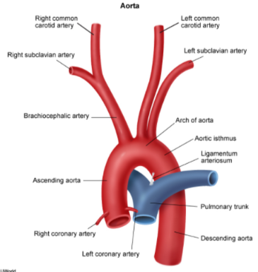
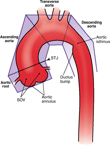
Aorta anatomy isthmus. The junction between the aortic arch and the descending aorta is called the aortic isthmus. Second during fetal life the isthmus area between left subclavian artery and the ductus arteriosus is narrow as this portion carries little blood. 9 superior mesenteric artery. Diameters of the abdominal aorta.
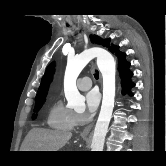
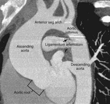
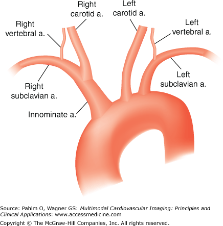
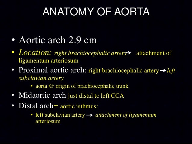
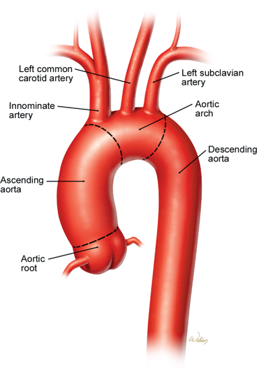
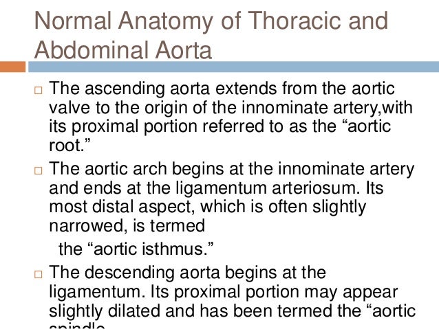
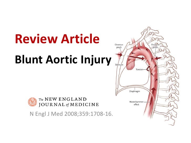
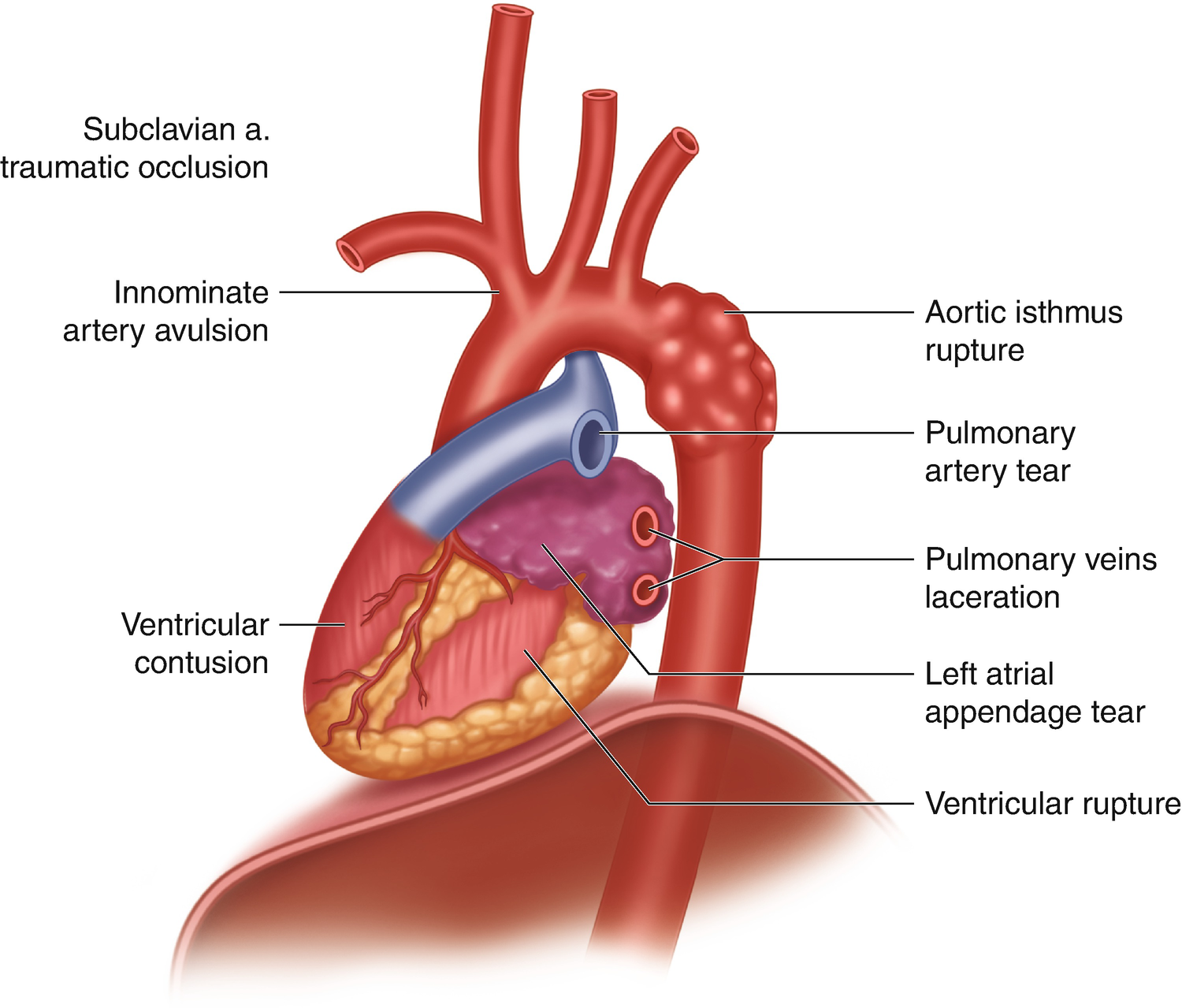
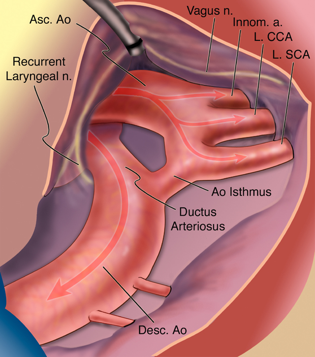
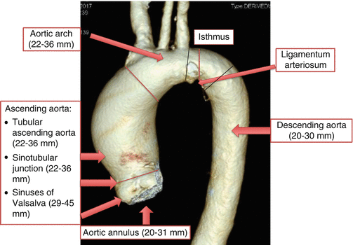
The aortic isthmus is the part of the aorta just distal to the origin of the left subclavian artery at the site of the ductus arteriosus. Isthmus aortae ta isthmus of aorta farlex partner medical dictionary c farlex 2012. The aorta distributes oxygenated blood to all parts of the body through the systemic circulation. The isthmus is a common site for coarctations and trauma.
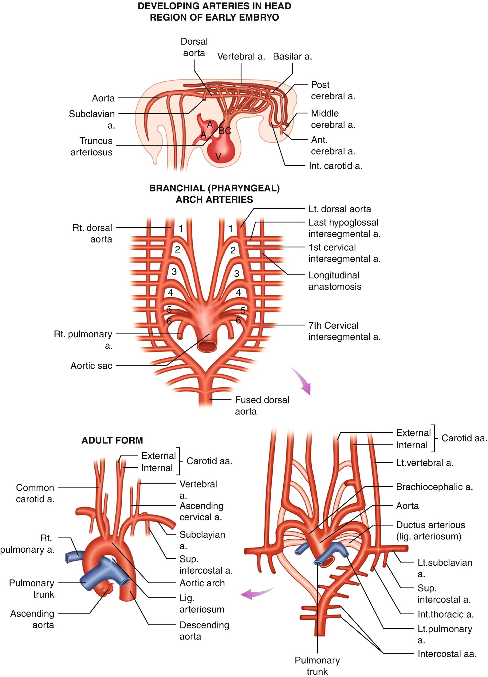
My cases submit a new case create a channel custom workshops my cases submit a new case. The heart pumps blood from the left ventricle into the aorta. It marks the partial separation of fetal blood flow derived from right and left ventricles. In the fetus this portion of the aorta is partly constricted because of the lack of flow within the aortic sac and ascending aorta.
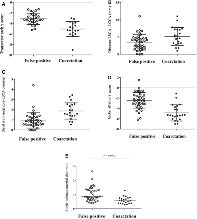
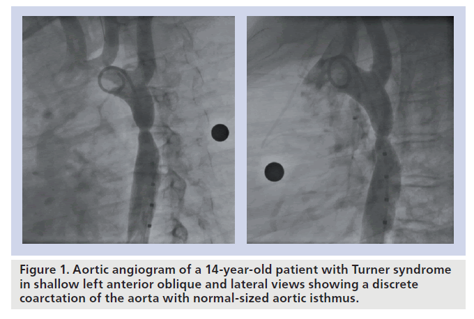
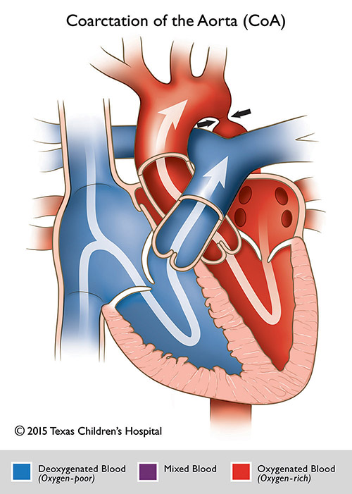
As the ductus arteriosus constricts after birth this tissue in isthmus area constricts causing development of coa 13 14. A slight constriction of the aorta immediately distal to the left subclavian artery at the point of attachment of the ductus arteriosus. The aorta begins at the top of the left ventricle the heart s muscular pumping chamber. The final section of the aortic arch is known as the isthmus of aorta.
This is so called because it is a narrowing isthmus of the aorta as a result of decreased blood flow when in foetal life. This portion of the aorta is partly constricted in the fetus because of the lack of flow within the aortic sac and ascending aorta. 243 035 cm. The descending aorta extends from the area.
Anatomy of the aorta. The aorta is the largest artery in the body. The aortic isthmus is a constriction of variable size of the aortic arch just distal to the origin of the left subclavian artery at the site of the ductus arteriosus behind the ligamentum arteriosum. This variant appearance may resemble a pseudoaneurysm of the aortic isthmus and therefore differentiation between the two is of particular importance.
Ductus diverticulum is most common at the aortic isthmus distal to the origin point of the left subclavian artery.












:background_color(FFFFFF):format(jpeg)/images/library/10023/Cadavar_Heart.png)

:background_color(FFFFFF):format(jpeg)/images/library/13386/T5ZCiKoPAprNRZN9BViZqg_Aorta.png)










:background_color(FFFFFF):format(jpeg)/images/library/13387/heart-in-situ_english.jpg)


